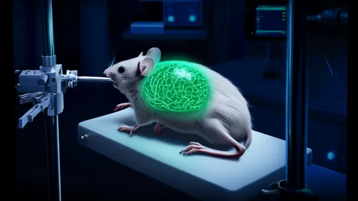
In a groundbreaking development that merges advanced materials science with neural imaging, researchers have unveiled a transparent cranial window technology enabling unprecedented long-term observation of brain activity. This innovation represents a significant leap forward from traditional methods that often required invasive procedures or provided limited temporal resolution. By creating a visually clear and biologically compatible interface, scientists can now monitor neural circuits with remarkable clarity over extended periods, opening new frontiers in understanding brain function and dysfunction.
The core breakthrough lies in the development of an optically transparent skull replacement that maintains full biocompatibility while allowing researchers to peer directly into the working brain. Unlike previous techniques that suffered from opacity issues or tissue rejection, this new approach utilizes specially engineered materials that integrate seamlessly with surrounding tissue. The window remains clear for months, permitting continuous observation of neural dynamics without the clouding or inflammation that plagued earlier iterations. This durability transforms how we study long-term processes like learning, memory formation, and neurological disease progression.
What makes this technology particularly revolutionary is its compatibility with multiple imaging modalities. Researchers can employ everything from two-photon microscopy to wide-field calcium imaging through the transparent window, capturing activity across entire cortical regions or zooming in on individual neurons. The stability of the preparation means that the same neural populations can be tracked day after day, revealing how circuits reorganize during experience-dependent plasticity. This longitudinal dimension provides insights impossible to obtain with acute experiments, showing not just how the brain works momentarily, but how it evolves over time.
The implications for neurological research are profound. For the first time, scientists can observe the real-time progression of conditions like Alzheimer's disease or epilepsy in animal models, watching as pathological changes unfold across neural networks. This could accelerate drug development by providing direct visual readouts of therapeutic efficacy at the circuit level. The technology also enables study of how the brain responds to injury and implements repair mechanisms, offering clues for treating trauma and stroke. Furthermore, it provides an ideal platform for testing neural prosthetics and brain-computer interfaces, allowing researchers to see how artificial inputs integrate with native circuitry.
Beyond disease research, the transparent window offers unprecedented access to fundamental neuroscience questions. How do neural representations form and stabilize during learning? What circuit mechanisms underlie decision-making? How do different brain regions coordinate during complex behaviors? These questions require observing the same neurons repeatedly during different experiences, exactly what this technology enables. Early studies using the technique have already revealed surprising dynamics in how neural ensembles compete and cooperate during task learning, overturning previous models based on snapshot observations.
The technological achievement extends beyond mere transparency. Researchers have engineered the window to minimize optical distortion while maintaining mechanical properties similar to natural bone, preventing stress on underlying tissue. Advanced coatings reduce biofilm formation and immune response, while nano-patterning techniques optimize light transmission for various wavelengths used in fluorescence imaging. Some versions even incorporate micro-electrode arrays or optical fibers for simultaneous manipulation and recording, creating truly multimodal platforms for interrogating neural function.
As with any transformative technology, challenges remain. The implantation procedure requires significant surgical expertise, though protocols are becoming standardized. There are also efforts to improve the material's long-term stability beyond the current several-month window and to enhance compatibility with increasingly sophisticated imaging techniques. Researchers are working on versions that could eventually be used in conjunction with MRI or PET scanning, combining the benefits of cellular resolution with whole-brain coverage.
Looking forward, this technology promises to bridge scales in neuroscience, connecting molecular changes to circuit-level phenomena and ultimately to behavior. It offers a powerful tool for studying the brain not as a static collection of cells, but as a dynamic system that continuously adapts and reorganizes. As the technology matures and becomes more widely adopted, it may well become the standard platform for systems neuroscience, much like patch clamping revolutionized cellular neuroscience decades ago.
The transparent cranial window represents more than just a technical improvement—it fundamentally changes what questions we can ask about the brain. By providing a permanent window into neural activity, it allows researchers to watch the brain's story unfold over time, rather than inferring plot from scattered snapshots. This continuous narrative will undoubtedly reveal surprises and complexities in how neural circuits support cognition, potentially transforming our understanding of both normal brain function and neurological disorders.

By /Aug 25, 2025

By /Aug 25, 2025

By /Aug 25, 2025

By /Aug 25, 2025

By /Aug 25, 2025
By /Aug 25, 2025

By /Aug 25, 2025

By /Aug 25, 2025
By /Aug 25, 2025
By /Aug 25, 2025

By /Aug 25, 2025

By /Aug 25, 2025

By /Aug 25, 2025

By /Aug 25, 2025

By /Aug 25, 2025
By /Aug 25, 2025

By /Aug 25, 2025

By /Aug 25, 2025
By /Aug 25, 2025

By /Aug 25, 2025